Retina (Medical)
What is the Retina?
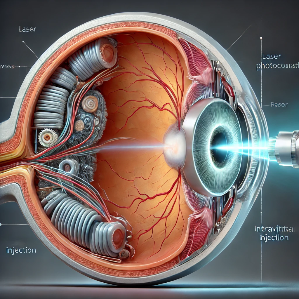
The retina is a thin layer of light-sensitive tissue located at the back of the eye. It plays a crucial role in vision by capturing light that enters the eye and converting it into electrical signals, which are then sent to the brain through the optic nerve. The brain interprets these signals to create the images we see. A healthy retina is essential for clear, sharp vision, as any damage to the retina can significantly impact your eyesight.
At Sanjeevani Netralaya, we specialize in the diagnosis and treatment of various retinal conditions, ensuring that your vision is protected and maintained with the highest standard of care.
What are Common Retinal Conditions?
Several retinal conditions can affect the clarity and quality of your vision. Some of the most common retinal disorders include:
1. Diabetic Retinopathy
- A condition that occurs when high blood sugar levels damage the blood vessels in the retina, causing them to leak or become blocked, leading to vision loss.
- Diabetic retinopathy is a common complication of diabetes and is one of the leading causes of blindness in adults.
2. Age-Related Macular Degeneration (AMD)
- AMD is a degenerative condition that affects the macula, the central part of the retina responsible for sharp, detailed vision.
- It can cause a gradual loss of central vision, making it difficult to read, recognize faces, or perform tasks that require detailed vision.
3. Retinal Detachment
- A serious condition in which the retina separates from the back of the eye, disrupting its blood supply and causing sudden vision loss.
- Retinal detachment is a medical emergency that requires prompt treatment to prevent permanent vision loss.
4. Retinal Vein Occlusion
- This occurs when a vein in the retina becomes blocked, leading to swelling, bleeding, and a reduction in vision.
- It is often associated with high blood pressure, diabetes, or other cardiovascular conditions.
5. Macular Hole
- A small break or tear in the macula that can cause blurred or distorted central vision.
- Macular holes are typically caused by aging, but they can also result from trauma or other eye conditions.
6. Epiretinal Membrane (Macular Pucker)
- A thin layer of scar tissue that forms on the surface of the retina, causing vision distortion or blurriness.
- The condition can develop with age or following eye surgery or injury.
Recognizing Symptoms of Retinal Conditions
Retinal diseases can manifest with a variety of symptoms, and early detection is critical to preventing permanent vision loss. Common symptoms to watch for include:
- Blurred or Distorted Vision: Difficulty seeing fine details or straight lines appearing wavy.
- Dark Spots or Floaters: Tiny specks or cobweb-like shapes drifting in your field of vision.
- Flashes of Light: Sudden flashes or flickering lights, especially in peripheral vision.
- Loss of Central Vision: Difficulty seeing objects in the center of your vision.
- Sudden Vision Loss: A rapid decrease in vision or a dark curtain-like effect across part of your vision.
If you experience any of these symptoms, it is important to seek immediate medical attention at Sanjeevani Netralaya.
Did You Know?
Many retinal conditions can be treated or managed effectively if detected early. Regular eye examinations are vital for early diagnosis and prevention of serious vision problems.
Diagnosis of Retinal Conditions
At Sanjeevani Netralaya, our experienced retinal specialists use advanced diagnostic tools and techniques to accurately diagnose retinal conditions. These may include:
- Ophthalmoscopy: A detailed examination of the retina using a specialized instrument called an ophthalmoscope.
- Optical Coherence Tomography (OCT): A non-invasive imaging test that provides cross-sectional images of the retina to detect abnormalities.
- Fluorescein Angiography: A procedure where a dye is injected into the bloodstream to highlight the blood vessels in the retina.
- Ultrasound Imaging: Used to examine the retina in cases where there is significant bleeding or clouding of the eye.
FAQs
How often should I have my retina checked?
It is recommended to have a comprehensive eye exam, including a retinal check, every 1-2 years, especially if you have risk factors such as diabetes or a family history of retinal conditions.
Can retinal conditions be prevented?
While not all retinal conditions can be prevented, managing risk factors like blood pressure, blood sugar levels, and cholesterol can reduce the likelihood of developing certain retinal diseases.
Is retinal surgery painful?
Retinal surgery is typically performed under local or general anesthesia, so patients do not feel pain during the procedure. Some discomfort or soreness may be experienced afterward, but this can be managed with medication.
What is the recovery time after retinal surgery?
Recovery time varies depending on the type of surgery and the patient’s overall health. Most patients can resume normal activities within a few weeks, but regular follow-up visits are essential to monitor healing.
